
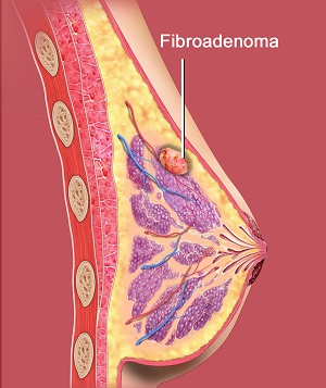
What is Fibroadenoma?
Fibroadenoma is a benign or noncancerous breast tumour that mostly occurs as a small, solid lump in the breast tissue measuring about 1 to 2 centimetres in size. Fibroadenomas are firm, smooth, rubbery, and round in shape that easily move around beneath the skin and do not cause any pain. They most commonly occur in young and premenopausal women, although women of any age can develop fibroadenoma.
Types of Fibroadenomas
Some of the types of fibroadenomas include:
- Simple Fibroadenoma: When you see this type of fibroadenoma under a microscope, it appears the same all over.
- Complex Fibroadenoma: This type of fibroadenoma mostly affects women of advanced age and has cells that grow at a greater rate.
- Juvenile Fibroadenoma: This is the most common type of breast lump seen in adolescents and girls between the age group of 10 to 18. It can grow large, but the majority shrink over a period of time and some even disappear.
- Giant fibroadenoma: This type of fibroadenoma can grow larger than 5 centimetres and may need to be excised if it replaces or presses on other breast tissue.
Causes of Fibroadenoma
It is unknown what exactly causes fibroadenoma. It is suspected to be linked to reproductive hormones as they occur more often at the time of puberty or pregnancy and go away post menopause. Use of hormone therapy is also suspected to be a potential cause of developing fibroadenoma.
Symptoms of Fibroadenoma
Fibroadenoma is usually painless and hence you may not notice it until you feel a lump beneath your breasts while you are bathing. Other times, your doctor may palpate it during your annual breast check-up or mammogram/ultrasound study. Unlike breast cancer, which can spread to other regions and grow larger in size, a fibroadenoma rests in the breast tissue without causing any symptoms such as swelling, redness, nipple discharge, or skin irritation around the breast.
Diagnosis of Fibroadenoma
In order to diagnose fibroadenoma, your doctor may perform the following diagnostic tests:
- A review of your medical history to check for any previous history of breast conditions
- A clinical breast examination to check for any lumps and other problems, such as nipple discharge or changes in the appearance of the breasts
- Mammogram – an X-ray examination of the breast to check for subtle changes not detected during a physical exam
- Ultrasound scan – use of high-frequency sound waves to create pictures of breast tissue to differentiate between a fluid-filled cyst and a solid mass
- Magnetic resonance imaging (MRI) – use of radio wave and strong magnetic field to produce detailed images of breast tissue to detect if the lump is cancerous or benign
- Breast biopsy – surgical removal of a sample of breast tissue or cells and fluid from a suspicious area for microscopic analysis
Treatment for Fibroadenoma
Some of the treatment approaches employed in the treatment of fibroadenoma include:
- Wait and Watch: If the fibroadenoma is small in size, your doctor mostly opts to simply wait and watch to see whether the lump shrinks or grows in size rather than try to excise it completely. This is the same method employed if fibroadenoma is noted during pregnancy or breastfeeding, where your doctor waits for your hormone levels to normalize to observe if the lump disappears by itself.
- Surgery: Your doctor may opt for surgical excision of the fibroadenoma if your clinical breast exam, imaging test, or biopsy confirms abnormality or if the fibroadenoma is deemed very large, increasing in size, or causing symptoms. Surgical procedures employed for the excision of fibroadenoma include:
- Excisional biopsy or lumpectomy: In this procedure, the lump is removed and sent for laboratory analysis to determine if the lump is cancerous or noncancerous in nature and whether it can grow and distort the surrounding breast tissue.
- Cryoablation: This is a minimally invasive surgical procedure, in which your doctor places a slim, wand-like instrument called a cryoprobe through your skin to create extremely cold temperatures using liquid nitrogen or argon gas to freeze and destroy the fibroadenoma.